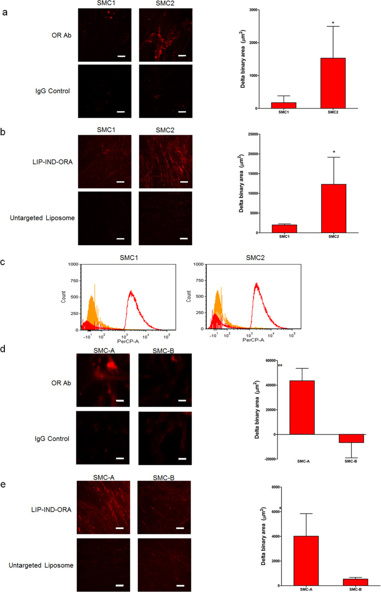
Figure 2. In vitro characterization of oxytocin receptor (OR) expression and correlated liposome targeting efficiency.
The in vitro experiments were conducted in triplicates in primary smooth muscle cells SMC1 and SMC2 isolated from pregnant mice (a–c), and in human smooth muscle cell lines SMC-A and SMC-B (d,e). OR expression was verified by immunofluorescence staining with OR antibody (OR-Ab, red) and analyzed via confocal microscopy (a,d), using IgG staining as a negative control. Liposome targeting specificity was analyzed via confocal microscopy (b,e), as well as via flow cytometry (c). All the images were analyzed and quantified using NIS-elements. Mean ± SEM, n = 9 images per sample. Scale bar = 50 μm. *p-value < 0.05, **p-value < 0.01 compared to IgG control (a,d) or to untargeted liposome (b,e).

