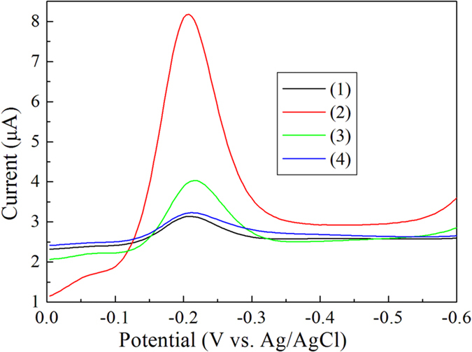
Figure 3. DPV of the antibody-aptamer sandwich assay in 0.1 M PBS (pH 7.4).

In the absence (1) and presence (2) of 20 nM Aβ oligomers after blocking of BSA. Curve 3 is the absence of 20 nM Aβ oligomers before blocking of BSA. Curve 4 is the presence of 20 nM Aβ oligomers without the immobilization of antibody.
