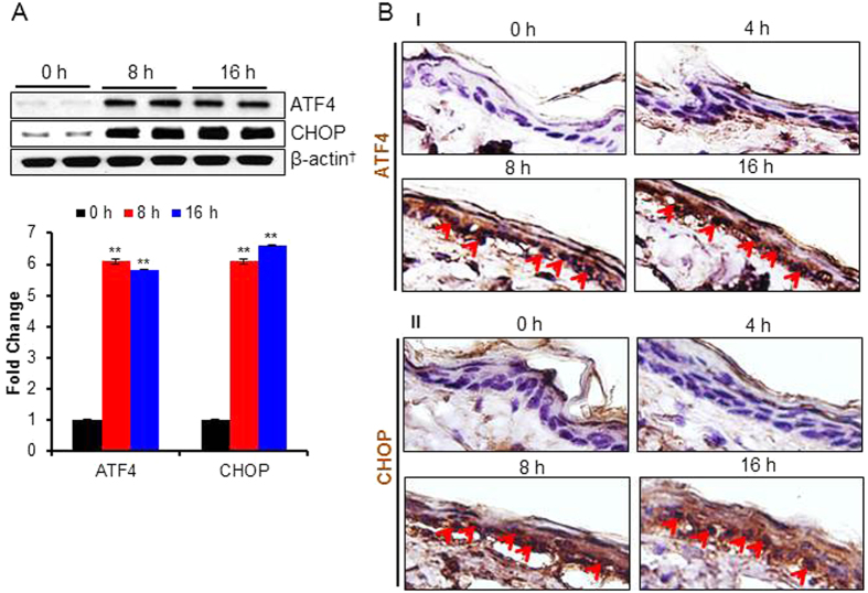
Figure 3. PAO-induces UPR signaling pathway in Ptch1+/−/SKH-1 mouse skin.
In this experiment, UPR signaling pathway markers were assessed in the skin of Ptch1+/−/SKH-1 mice treated either with vehicle or PAO (100 μg/mouse) for different time intervals (0–16 h). (A) Western blot analysis of ATF4 and CHOP in the skin tissue lysate of PAO-treated samples at 0 h, 8 h and 16 h. Histogram showing dentiometric analysis of western blots. (B) Immunohistochemical staining of ATF4 and CHOP. Arrows showing expression and nuclear translocation of ATF4 (B-I) and CHOP (B-II) in the PAO-treated skin sections.

