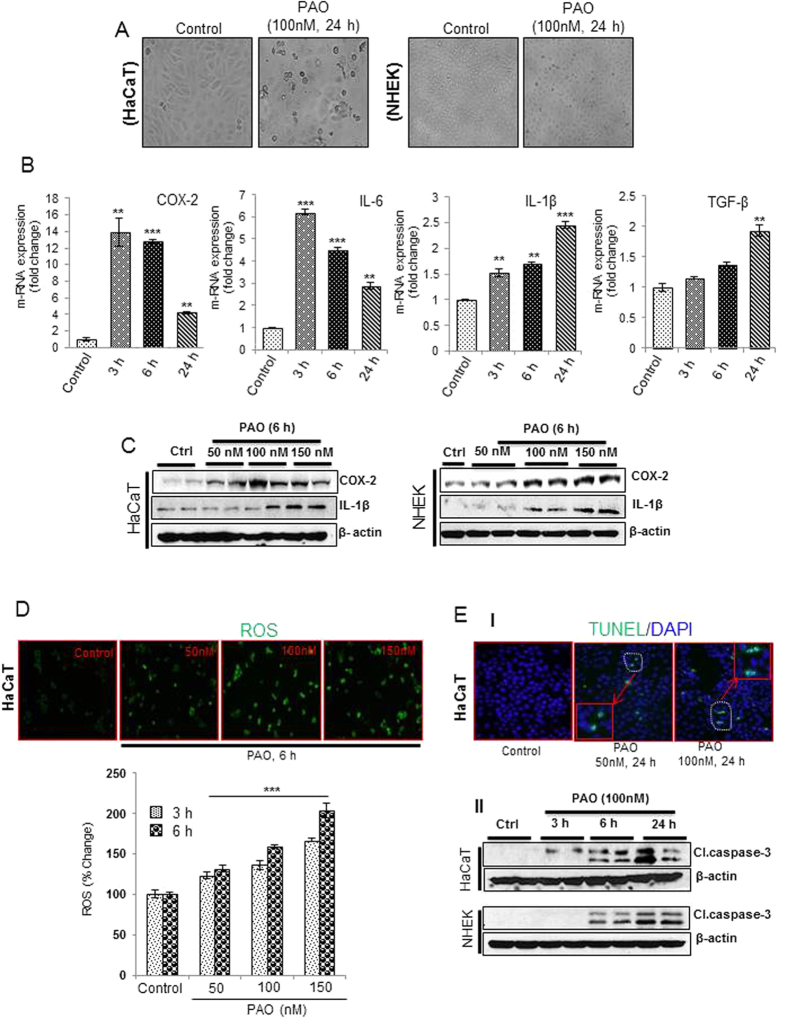
Figure 5. Dose- and time-dependent kinetics of cytokines production, ROS generation and apoptosis induction in PAO-treated human keratinocytes.
In these experiments, human skin keratinocytes (HaCaT/NHEK) were treated with PAO (50–150 nM) for 3–24 h time intervals. (A) Phenotypic alterations including cell rounding, loss of cell adhesion and blebbing in HaCaT and NHEK in culture. (B) Real time PCR showing time-dependent response for pro-inflammatory cytokines (IL-6, IL-1β & TGF-β) and COX-2 expression. (C) Western blot analysis showing dose-dependent effects of PAO on the expression level of IL-1β and COX-2 in HaCaT and NHEK. (D) Fluorescence-based microphotographs and ELISA-based plate reader assays showing dose and time-dependent effects of PAO on ROS production in HaCaT cells. (E-I) Microphotographs of fluorescent TUNEL-positive cells at various doses of PAO. (E-II) Time-dependent effects of PAO on western blot analysis of cleaved caspase-3 in HaCaT and NHEK. Data are expressed as mean ± SEM. **P < 0.01 and ***P < 0.001 values show significance levels when compared to control.

