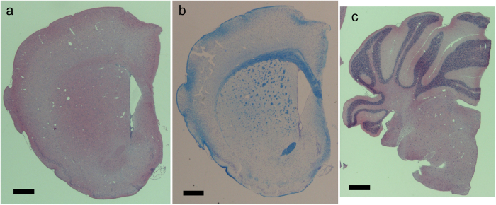
Figure 2. Paraffin sections of right hemisphere of murine brain.
Sections 2 μm in thickness were prepared for hematoxylin-eosin staining, and 4 μm sections were prepared for Klüver-Barrera staining. Scale bars: 500 μm. (a) A hematoxylin-eosin section of cerebrum. The anterior commissure and corpus callosum appear as eosin-stained (pink) structures. (b) A Klüver-Barrera section of cerebrum. Myelinated axonal tracts in the anterior commissure, corpus callosum, and striatum appear as blue structures. (c) A hematoxylin-eosin section of cerebellum. Granular layers appear as hematoxylin-stained (purple) structures.

