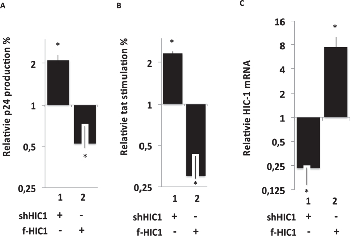
Figure 4. Effect of HIC1 over-expression or knock-down on HIV-1 in microglial cells.
Microglial cells were transfected either with the pNL-4.3 provirus (A) or the episomal pLTR-luciferase reporter (B) and the indicated plasmids. 48-hours later, supernatants were harvested (A) or cells were lysed (B). Viral p24 concentrations were titrated in supernatant and normalized relatively to their appropriate empty vector control (A). Cell lysates were subjected to luciferase assay, normalized with the renilla luciferase system and expressed as relative Tat stimulation with their respective control (B). Nuclear cell extracts were obtained after 48-hours transfection of the indicated vectors and subjected to western-blot experiments to detect endogenous knock down and over-expressed HIC1 (respectively lane 1,2 and 3). As a control we checked the relative HIC1 mRNA following a HIC1 knock down (shHIC1) or following overexpression of flag-HIC1 (f-HIC1) (C).

