
Figure 5. HIC1 relocates TAT in sub-nuclear ball like structures in which they interact.
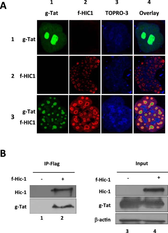
(A) Microglial cells were transfected with pGFP-TAT (row 1), pFlag-HIC1 (row 2) or both (row 3). 48-hours post-transfection cells were fixed and stained with TOPRO-3 (column 3) to detect the nucleus. Flag-HIC1 was detected by incubating cells with an anti-flag antibody and immunostained with a cyanine 3-labelled secondary antibody (column 2). Localization of Flag-HIC1 and GFP-TAT has been acquired by confocal microscopy. (B) HEK293T cells were transfected with pGFP-TAT alone or in combination with pFlag-HIC1. 48-hours later cells were lysed and nuclear extracts have been subjected to immunoprecipitation with an anti-Flag tag antibody. TAT and HIC1 were detected by western blot with respectively anti-TAT and anti-Flag antibodies.