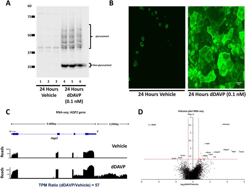
Figure 1. The V2-receptor-specific vasopressin analog dDAVP (0.1nM for 24 hours) increases the abundance of aquaporin-2 (AQP2) protein and mRNA in mouse collecting duct cells (mpkCCD).
(A) Western blot shows large increase in AQP2 protein (Vehicle, Lanes 1, 2 and 3; dDAVP, Lanes 4, 5 and 6). (B) Confocal images of immunofluorescence immunocytochemistry for AQP2 confirms the large increase in AQP2 protein abundance in response to dDAVP. Scale, 15 μm. (C) RNA-seq data for Aqp2 gene shows large increase in AQP2 mRNA. RNA-seq reads for a single pair of samples are mapped to exon-intron structure of gene for vehicle-treated cells (above) and dDAVP-treated cells (below). Most reads are mapped to 3′-most exon because reverse transcripton was primed with oligo-dT. (Supplemental Fig. 1 shows the same mapping on a linear scale.). (D) Volcano plot for RNA-seq data for all 8393 detectable transcripts shows that a relatively small fraction of transcripts are regulated by vasopressin, but that AQP2 mRNA is increased by a relatively large amount. The horizontal axis shows the mean log2(dDAVP/Vehicle) for 9 pairs of samples. The vertical axis shows −log10P for t-tests for each gene. Vertical dashed lines show 95% confidence interval for random variation based on Vehicle:Vehicle comparisons (2 × SD). The use of two selection factors represented on x- and y- axes provides stringent identification of vasopressin-responsive transcripts in upper right (red points) and upper left (blue points).

