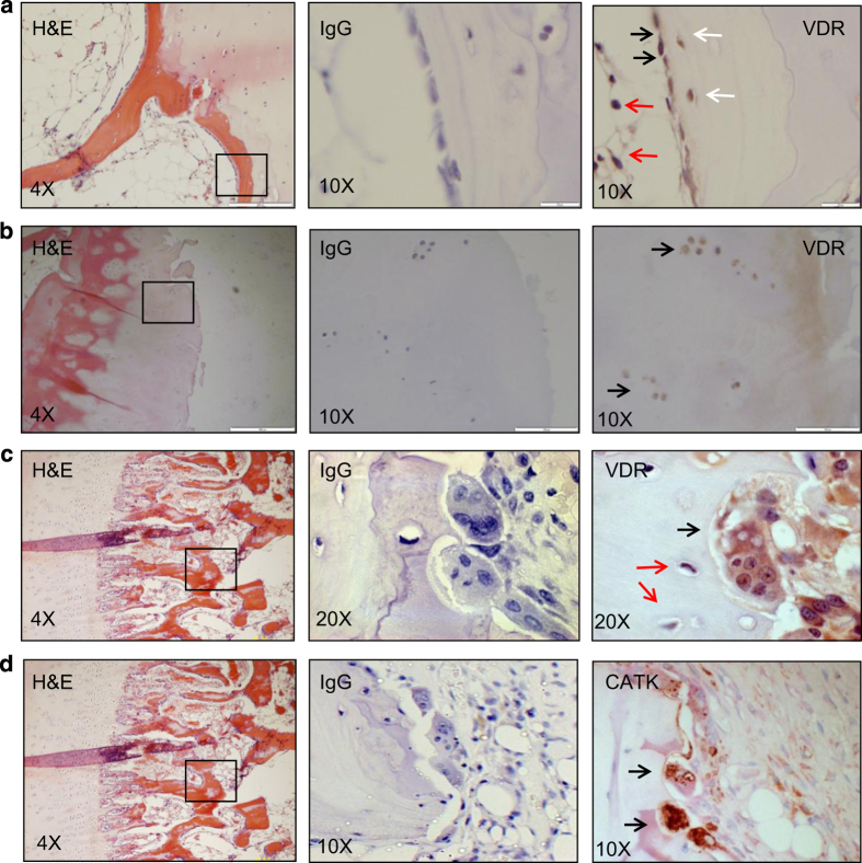
Figure 1.
VDR is expressed by BMSCs, osteoblasts, osteocytes, chondrocytes, and osteoclasts in bone tissue. Serial sections of human bone samples, right column; hematoxylin and eosin staining, middle column; negative staining (IgG), left column; IHC of VDR (a–c), and (d) Cathepsin K (CATK). Positive cross-reactivity of VDR was observed in bone lining osteoblasts (black arrows in a), newly embedded osteocytes (white arrows in a), adipocytes (red arrows in a), chondrocytes (black arrows in b), bone resorbing multinucleated osteoclast (black arrow in c). Negative cross-reactivity was observed in mature osteocytes (red arrows in c).

