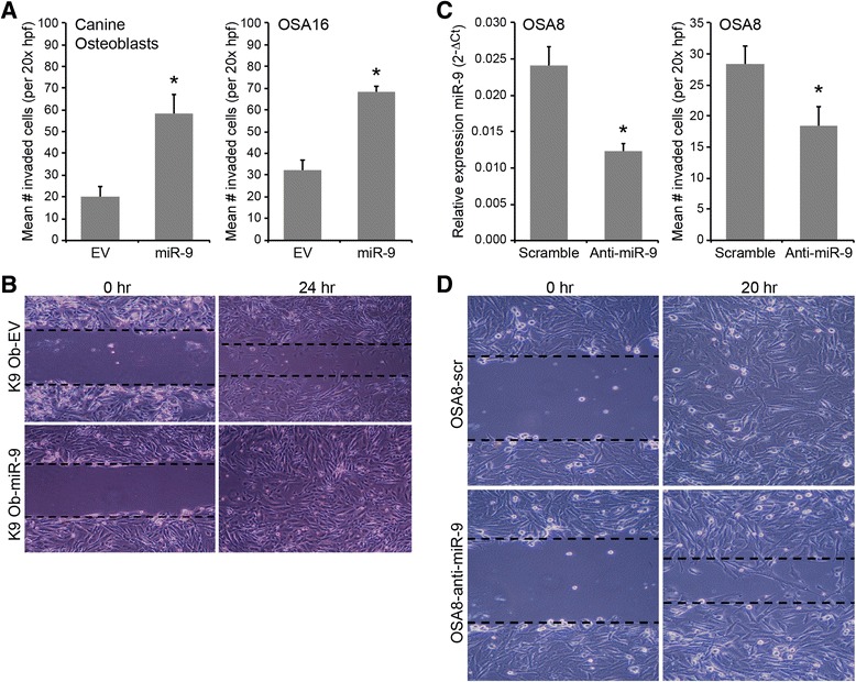
Fig. 5.

MiR-9 enhances invasion and migration in normal canine osteoblasts and OS cell lines. a The invasive capacity of normal canine osteoblasts or OSA16 cells transduced with either empty vector or pre-miR-9-3 lentivirus was evaluated using standard Matrigel invasion assays. Cells (5 × 10⁴) were plated in serum free medium and transferred onto cell culture inserts coated with Matrigel® for 24 h. After incubation, cells remaining on the upper surface of the insert membrane were wiped away using a cotton swab, and cells that had migrated to the lower surface were stained with crystal violet and counted in ten independent 20x hpf for each sample. Three independent experiments were performed and all assays were performed in triplicate wells (Bars: SD. Statistical analysis: one-way ANOVA, *p < 0.001). b Cell migration was assessed in canine osteoblasts transduced with either empty vector or pre-miR-9-3 lentivirus using standard wound-healing assays. Cells were seeded in complete medium and grown until confluent in 6-well plates. A gap was created using a P200 pipette tip, and medium was replaced with serum-free medium. After 24 h, cell migration was evaluated by digital photography. c Canine OSA8 cells were transduced with miRZip-9 (anti-miR-9) or scramble vector and miR-9 levels were assessed by real-time PCR to confirm transduction efficiency (*p < 0.0009). Cell invasion was assessed in canine OSA8 cells transduced with scramble or miRZip-9 (anti-miR-9) lentivirus using standard Matrigel invasion assays as described above. Three independent experiments were performed and all assays were performed in triplicate wells (*p < 0.0002). d Cell migration was assessed in OSA8 cells transduced with miRZip-9 (anti-miR-9) or scramble vector using standard wound-healing assays as described above. After 20 h, cell migration was evaluated by digital photography
