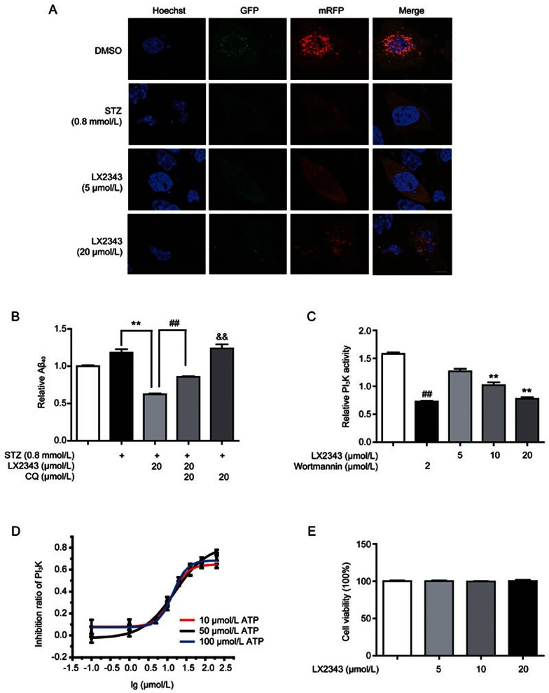
Figure 6.
LX2343 as a PI3K inhibitor stimulated autophagy in the promotion of Aβ clearance. CLSM images of SH-SY5Y cells transiently expressing mRFP-GFP-LC3 (A) (green and red puncta indicate GFP and mRFP, respectively. Scale bar: 5 μm, n=3). CQ-based ELISA result demonstrated that CQ enhanced Aβ levels and partially reversed LX2343-induced Aβ reduction in SH-SY5Y cells (B) (t test, **P<0.01 vs STZ; ##P<0.01 vs STZ combined with LX2343; &&P<0.01 vs DMSO). LX2343 dose-dependently inhibited PI3K activity in vitro (C) (wortmannin: PI3K inhibitor. One-way ANOVA, Dunnett's multiple comparison test. n=3. **P<0.01 vs DMSO; wortmannin, t test, n=3). LX2343 dose-dependently inhibited PI3K in the presence of the indicated concentrations of ATP (D). In the presence of 10 μmol/L of ATP, the IC50 of LX2343 is 13.11±1.47 μmol/L, in the presence of 50 μmol/L ATP, the IC50 of LX2343 is 13.86±1.12 μmol/L, in the presence of 100 μmol/L ATP, the IC50 of LX2343 is 15.99±3.23 μmol/L. LX2343 concentration was expressed in log10 scale. MTT assay result demonstrated that LX2343 had no effects on cell viability in SH-SY5Y (E) (one-way ANOVA, Dunnett's multiple comparison test, n=3). GAPDH was used as loading control in Western blot assays. All data were obtained from three independent experiments and presented as mean±SEM.

