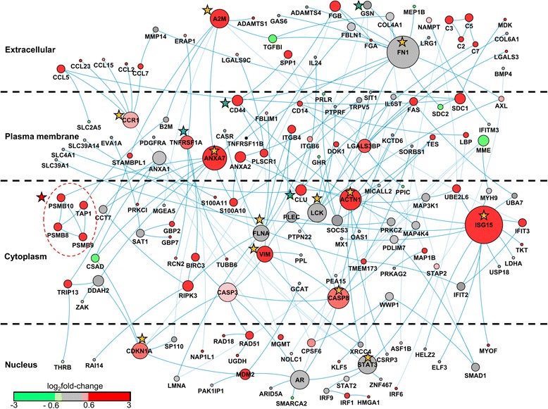
Fig. 6.

Acute kidney injury (AKI)-relevant human protein-protein interaction sub-network. The protein nodes are distributed according to cellular localization. The size of the node represents the number of connections in the sub-network. The nodes are colored according to the average log2 fold-change ratio in chemical exposures that cause kidney necrosis. Proteins encoded by genes with average log2 fold-change ratios greater than 0.6 are shown in red. Proteins encoded by genes with average log2 fold-change ratios between 0.6 and −0.6 are shown in grey. Proteins encoded by genes with average log2 fold-change ratios less than −0.6 are shown in green. Orange stars denote hub proteins with >5 connections, green stars denote non-hub proteins with a high betweenness centrality (>0.09). The red star and dotted circle identify the highest interconnected region of the network associated with the immunoproteasome
