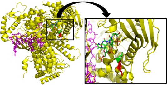
Figure 6.

Superposition of yeast DNA polymerase and ssDNA substrate from Escherichia coli DNA polymerase Klenow fragment. Yeast DNA polymerase epsilon (yellow) and bound dsDNA (magenta) with ssDNA substrate (red, green, blue, orange) from E. coli DNA polymerase Klenow fragment superimposed onto the exonuclease domain of the yeast DNA polymerase. The magnification shows a slice through the polE structure (4M8O.pdb) superimposed with the ssDNA (red, green, blue, orange) in the exo‐site from the structure of the Klenow fragment (18Y.pdb). The side chains of the Lys425 and the Leu424 residues are shown in red and green, respectively. [Color figure can be viewed in the online issue, which is available at wileyonlinelibrary.com.]
