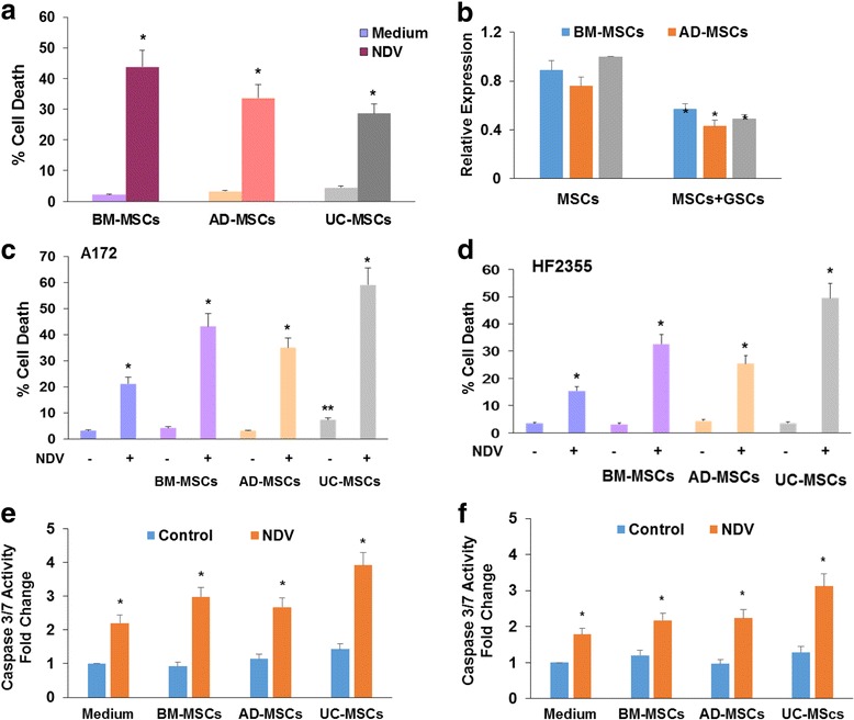
Fig. 2.

Conditioned medium of NDV-infected MSCs exerts potent cytotoxic effects on glioma cells and GSCs. MSCs derived from BM, AD or UC tissue were infected with NDV (5 MOI) and cell death was determined after 3 days using LDH assay (a). MSCs were infected with NDV (2 MOI), washed three times and co-cultured with A172 cells in transwell plates with 0.4 μm for 48 h. The A172 cells were washed three times and the presence of NDV in the cells was determined using RT-PCR (b). The A172 cells (c, e) or HF2355 GSCs (d, f) were either infected with 2 MOI NDV or incubated with medium conditioned from control or NDV-infected MSCs for 2 days. Cell death was determined using LDH assay (c, d) or caspase 3/7 activity assay (e, f) after 24 h. The results are presented as means ± SE and represent three different experiments. * p < 0.001 (control vs. infected cells). AD adipose tissue, BM bone marrow, GSC glioma stem cell, MSC mesenchymal stromal cells cell, NDV Newcastle disease virus, NSC neural stem cell, UC umbilical cord
