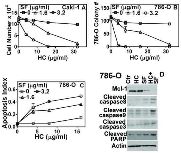
Figure 1. Effect of HC, SF and HC+SF on RCC cells.
A: Caki-1 cell viability was determined after 72-h exposure to HC, SF or HC+SF at indicated concentrations. Live cells were counted; B: Clonogenic survival of 786-O cells was determined after 7-days. C: Apoptosis was measured following the exposure of 786-O cells to HC, SF or HC+SF for 72 h. Y-axis: O.D. 405 nm. Data for A–C: Mean±sd. D: Immunoblotting of 786-O cells for apoptosis indicators following 48-h exposure to HC (16-μg/ml), SF (3.2-μg/ml) and HC+SF.

