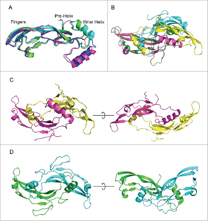
Figure 3.

Comparison of myostatin with PDB crystal structures. (A) myostatin from the humanized (blue) and chimeric (magenta) co-crystal structures are aligned with myostatin from PDBID:3hh2 (green) and PDBID3sek (cyan). The pre-helix and wrist helix are labeled for myostatin from the chimeric and humanized structures. These are shifted downwards for the 3hh2 and 3sek structures. (B) Alignment of dimeric myostatin from chimeric structure (magenta and yellow) with 3hh2 (green and cyan). (C) Linear structure of dimeric myostatin from the chimeric crystal structure. (D) Butterfly conformation of the dimeric myostatin from the 3hh2 crystal structure.
