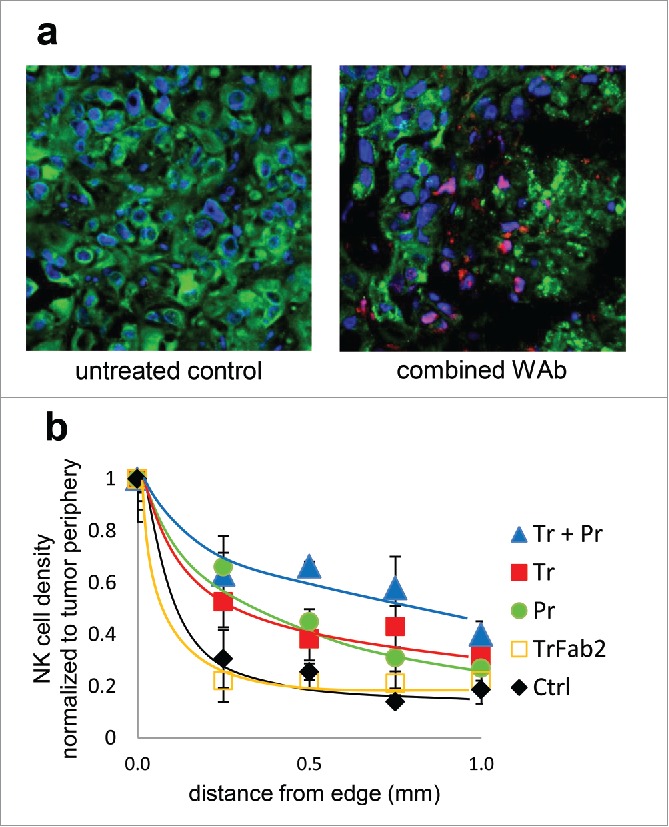
Figure 5.

NK cell infiltration of JIMT-1 tumor xenografts. Frozen sections of excised tumors were fluorescently labeled for HER2 (green), NK cells (CD45, red/magenta), and nucleic DNA (blue). Sections from an untreated control (left) and one treated with trastuzumab plus pertuzumab combination (right) are shown in (a). Treatment with antibodies enhances NK cell invasion, which results in extensive tumor lesion and a less dense tumor tissue structure. NK cell penetration into the tumor was quantitatively analyzed by counting CD45+ nucleated cells in 0.05 mm2 areas starting from the side of the tumor toward the center (b).
