Figure 4.

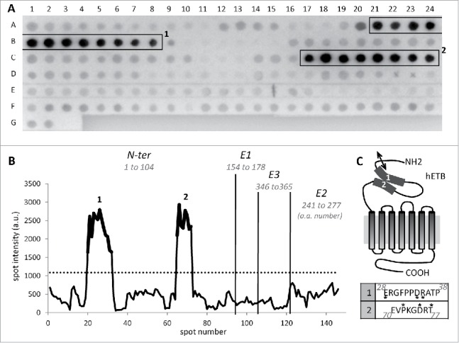
Rendomab-B4 recognizes a discontinuous epitope on hETB N-terminal tail. (A) To determine rendomab-B4 epitope, a Pepscan membrane spotted with dodecapeptides corresponding to extracellular domain and loops of the ETB receptor, was used. Hybridized monoclonal antibody was revealed using an alkaline phosphatase conjugated secondary antibody. The highest consecutive reactive spots are in boxes 1 and 2. Each spot signal obtained on the membrane was quantified. (B) The profile of hybridization of rendomab-B4, corresponding to the spot intensity as a function of the spot number on the membrane, was drawn. Vertical lines symbolize the separation between N-terminal (N-ter) domain and extracellular loops (E1, E2 and E3) of the receptor. The numbers of the amino acids at the extremities of each domain are indicated in italics. The dotted line corresponds to 3 times the background calculated as the mean intensity of all the spots except those in boxes. Parts of the curve in bold print (1 and 2) correspond to spots in boxes drawn in A. (C) Schematic representation of the human ETB and putative localization of the rendomab-B4 regions of recognition (1 and 2). The double arrow symbolizes the cleavage site of ETB signal peptide. Corresponding amino acid sequences of regions 1 and 2 are indicated in the table below. Amino acid noted * are not conserved between human and rodent (rat and mouse) ETB. This experiment was done twice with 2 independent membranes. Isotype control antibody generated no staining (data not shown).
