
Figure 4.
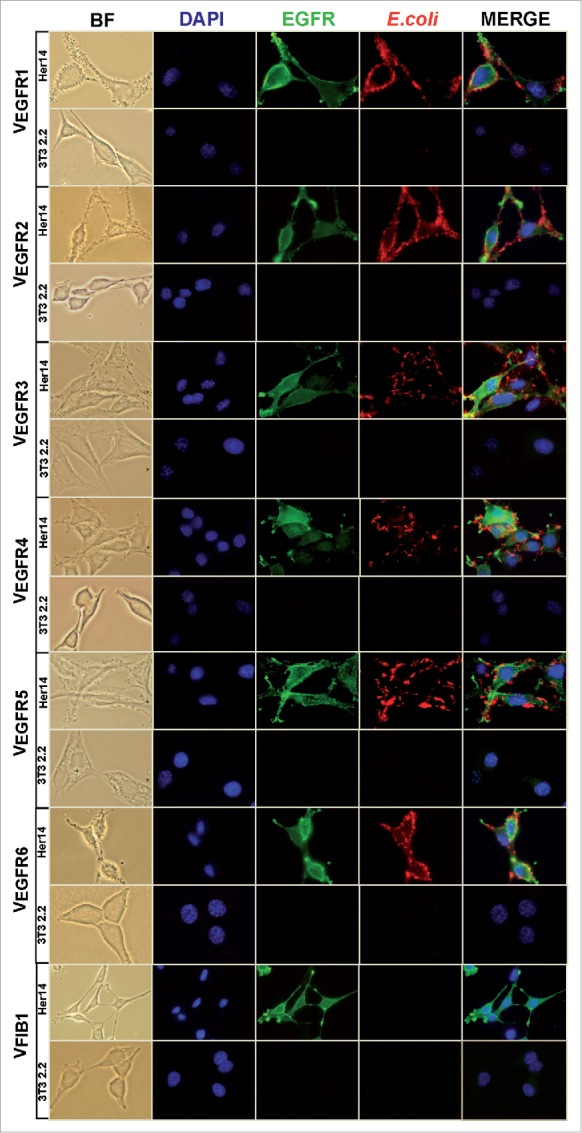
Adhesion to tumor cells of E. coli bacteria displaying the selected anti-EGFR Nbs. Bright field (BF) and fluorescence microscopy images of Her14 (EGFR+) and NIH-3T3 2.2 (EGFR-) cells grown in culture and infected with E. coli bacteria displaying the indicated Nb clone. Microscopy images showing the specific adhesion of bacteria with selected anti-EGFR Nbs to Her14 cells, but not to NIH-3T3 2.2 cells. Also included is an E. coli bacterial clone displaying a non-relevant Nb (VFIB1). Bacteria were labeled with anti-E. coli polyclonal Ab (red), EGFR was labeled with anti-EGFR mAb (green), DNA and cell nuclei were labeled with DAPI (blue).