
Figure 2. Hinokitiol induces autophagy in neuronal cells.
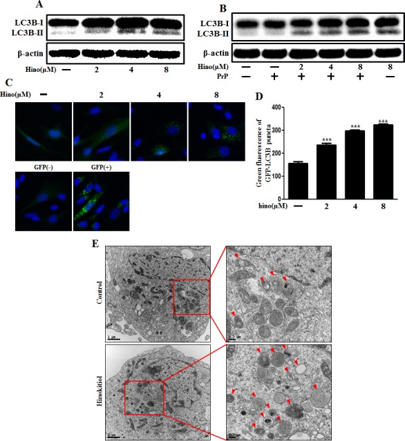
The primary neurons were treated with 2, 4, and 8 μM of hinokitiol for 6 h A. and were then exposed to 100 μM PrP (106-126) for 6 h B.. The treated cells were assessed for LC3B production by Western blot analysis. SK-N-SH cells were mixed with a titration (30MOI) of BacMam GFP-LC3B virus over 18 h and were then treated with hinokitiol in a dose-dependent manner for 6 h C., D.. Negative control reagent and positive control reagent (CQ) at the same time. E. SK-N-SH cells were incubated with 8 μM of hinokitiol for 6 h and analyzed by TEM. Arrowheads indicate autophagosomes. *** p < 0.001; significant differences when compared with control and each treatment group. Hino, hinokitiol; PrP, Prion peptide (106-126); GFP (+), Positive control; GFP (−), Negative control.