
Figure 7. Hinokitiol-induced autophagy protects cytotoxicity via Hif-1α stabilization.
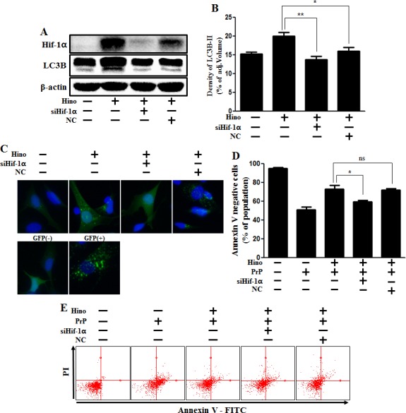
A. Hif-1α small interfering RNA (siHif-1α)-transfected or negative control siRNA (NC)-transfected SK-N-SH cells were incubated with 8 μM of hinokitiol for 6 h. Western blots for Hif-1α, LC3B proteins were analyzed from SK-N-SH cells. B. Bar graph indicating the averages of LC3B-II levels. C. SK-N-SH cells were mixed with a titration (30MOI) of BacMam GFP-LC3B virus over 18 h and were then treated with hinokitiol for 6 h, Negative control reagent and positive control reagent (CQ) at the same time. D. Bar graph of cell viability indicating the averages of annexin V-negative cells. E. Cell viability was measured by Annexin V assay from siRNA-treated cells. *p < 0.05; significant differences when compared with control and each treatment group. Hino, hinokitiol; Prion peptide (106-126); Hif-1α, Hypoxia-inducible factor-1 alpha; p-Akt, phosphorylation of Akt; NC, Negative control.