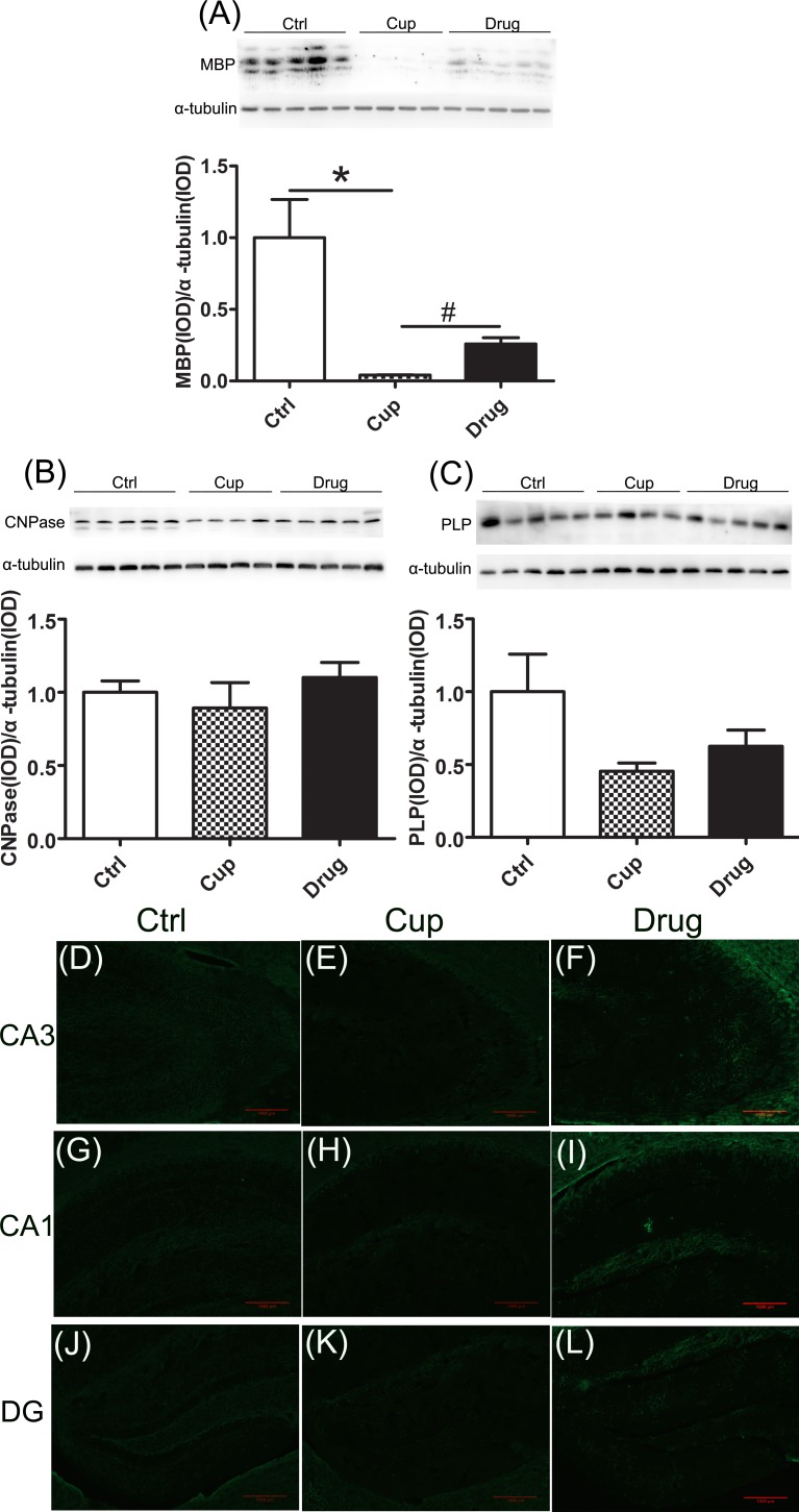
Figure 2. Demyelination in the hippocampus.
Myelin associated proteins in the hippocampus, including MBP A., CNPase B. and PLP C., were detected among the three groups using Western-blot. Figures D.-L. were displayed the immunofluorescence staining of MBP in subregions of the hippocampus, including Ca3 (D-F), Ca1 (G-I) and DG (J-L). *denotes statistical significance compared with controls (P < 0.05). #denotes statistical significance compared with the cuprizine-fed mice without treatment (P < 0.05). Images were captured from stained frozen sections using a fluorescence microscope equipped with 10×objectives. Scale bar, 1000μm.

