
Figure 1. Baseline levels of CD4+ T cells are increased in lungs and spleens of aged mice.
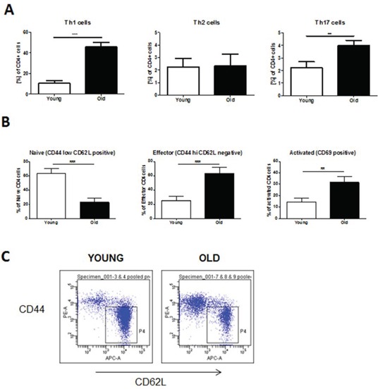
For flow cytometric analysis single-cell suspensions were prepared from whole lung tissue or spleens of young (2 months of age) and old (12 months of age) mice. Surface marker and intracellular cytokine stainings were subsequently performed. A. Th1 cells were defined by expression of CD4 and IFNγ, Th2 cells by expression of CD4 and IL-4, and Th17 cells by expression of CD4 and IL-17A. B. CD4+ effector and activated T cells were characterized by expression of CD44 and CD69, respectively; C. Representative FACS plots of stainings for CD4+ effector T cells. Data were combined from 2 independent experiments with n = 4 and are given as mean values ± SD; one-way ANOVA following Bonferroni post test with *p < 0.05, **p < 0.01, ***p < 0.001.