
Figure 2. Aged mice develop emphysema and airway remodeling after 3 months of CS exposure.
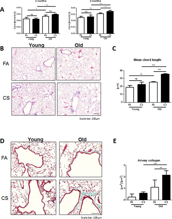
A. Lung function measurements for dynamic lung compliance were performed in intubated animals after 2 months of CS exposure and in tracheostomized animals after 3 months of CS exposure. B. Representative micrographs of HE-stained lung tissue sections from young and old FA and CS-exposed mice; scale bar 200 μm. C. Quantitative measurement of emphysema was determined by design-based stereology of HE-stained lung tissue sections using an Olympus BX51 light microscope equipped with the computer-assisted stereological toolbox newCAST. D. Representative micrographs of Masson's Trichrome staining of lung tissue sections from young and old FA and CS-exposed mice; scale bar 100 μm. E. Total volume of airway collagen per basal membrane was determined via quantitative morphological assessment. Data were combined from 2 independent experiments with n = 8 and are given as mean values ± SD; one-way ANOVA following Bonferroni post test with **p < 0.01, ***p < 0.001.