
Figure 5. Macrophage markers are increased in the lungs of CS-exposed aged mice.
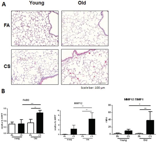
A. Representative micrographs of immunohistochemical stainings for MMP12 in lung sections from FA control and CS-exposed young and old mice; scale bar 100 μm. B. Lung tissue mRNA expression of macrophage markers F4/80 and MMP12 as well as the ratio of MMP12/TIMP1 was measured by qPCR. Data were combined from 2 independent experiments with n = 8 and are given as mean values ± SD; one-way ANOVA following Bonferroni post test with *p < 0.05, **p < 0.01, ***p < 0.001.