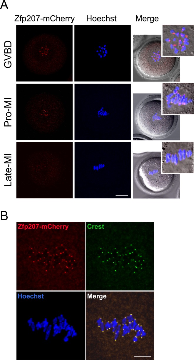
Figure 1. Localization of Zfp207 during mouse meiotic maturation.
A. cRNA of Zfp207-mCherry was microinjected into GV oocytes which were then cultured to various developmental stages. mCherry signals were acquired under the confocal microscope at 594 nm laser. Chromosomes were counterstained with Hoechst. GVBD, oocytes at germinal vesicle breakdown stage; Pro-MI, oocytes at first prometaphase stage; Late-MI, oocytes at late stage of first metaphase. Scale bar, 20μm. B. Zfp207-mCherry expressing oocytes were immunostained with kinetochore marker Crest and then counterstained with Hoechst. Scale bar, 5μm.

