
Figure 4. Depletion of Zfp207 disrupts kinetochore-microtubule attachment in mouse oocytes.
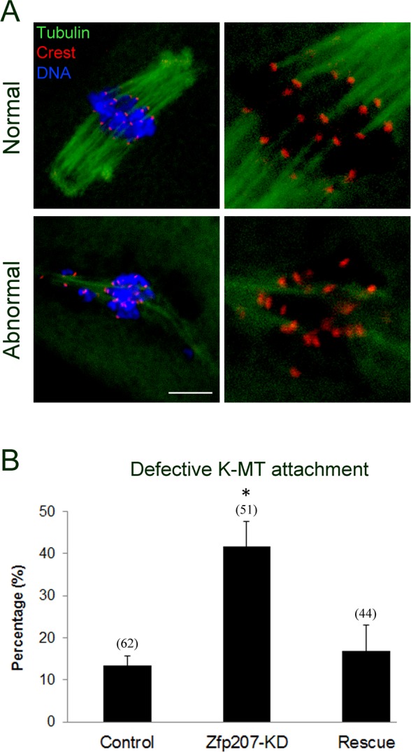
A. Representative images of normal and abnormal kinetochore-microtubule attachment in mouse oocytes. Oocytes were immnunostained with α-tubulin-FITC antibody to visualize spindle, with Crest to visualize kinetochore, and counterstained with Hoechst to visualize chromosome. Scale bar, 10μm. B. The rate of defective kinetochore-microtubule attachment was recorded in the control, Zfp207-KD and rescue oocytes. Data were presented as mean percentage (mean ± SEM) of at least three independent experiments. Asterisk denotes statistical difference at a p < 0.05 level of significance.