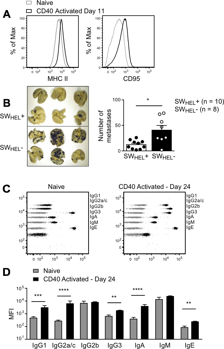
Figure 7. Effect of anti-CD40 activated B cells and isotype switched anti-tumor antibody in a lung metastasis model.

Rag2−/− SWHEL+ and Rag2−/− SWHEL- mice were injected i.v. with 1×10⁶ tumor cells followed by i.p. anti-CD40 on days 3 and 6. A. Splenocytes from two anti-CD40 treated Rag2−/− SWHEL+ mice 11 days after tumor challenge were pooled and stained with B cell activation markers, as indicated. Black histograms: anti-CD40 activated Rag2−/− SWHEL+ B cells; grey histograms: naive Rag2−/− SWHEL+ mice. Additional mice were euthanized on day 24, lungs were collected to count metastatic foci and serum was sampled to measure antibody concentration. B. Left: Representative photos of lungs from Rag2−/− SWHEL+ and Rag2−/− SWHEL- mice. Right: Number of metastases. Each dot represents an individual animal, with the bar representing mean±SEM (SWHEL+: n = 10, SWHEL-: n = 8). * = P < 0.05 by nonparametric Mann-Whitney test. C. Representative dot plots depicting antibody isotype CBA data from Rag2−/− SWHEL+ mice (black) overlaid on control CBA plot (grey). Left panel: naïve Rag2−/− SWHEL+ mouse. Right panel: anti-CD40 treated Rag2−/− SWHEL+ mouse after i.v. tumor challenge (time of death: Day 24). D. MFI of CBA signals for each isotype before tumor challenge and on day of death, mean±SEM, n≥9/group. * = P < 0.05, ** = P < 0.01, *** = P < 0.001, **** = P < 0.0001 by unpaired Student's t test.
