
Figure 3. HP1α directs the localization of PC2 and SENP7L.
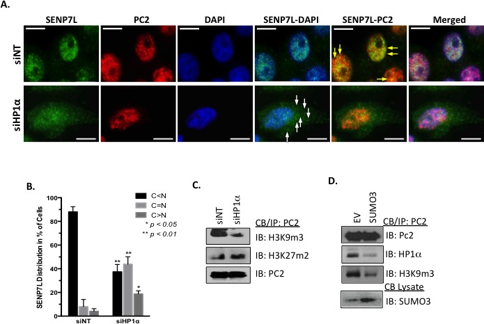
A.-B. HP1α loss alters SENP7L subcellular localization. Cells were incubated with either non-targeting (siNT) or HP1α-specific (siHP1α) siRNA for 48 hr. Immunofluorescence was performed with SENP7L and PC2 antibodies and nuclei stained with DAPI. Total cells and SENP7L distribution was evaluated in randomly selected 20x magnification fields (siNT: n = 3 and siHP1α: n = 7); percent of cells with cytosolic (C) versus nuclear (N) SENP7L were analyzed using ANOVA and Tukey's post-hoc. C. PC2's enrichment at H3K9m3 requires HP1α. Chromatin-bound PC2 was isolated from MCF7 cells treated with either siNT or siHP1 as described above. Western blot analysis was performed to evaluate PC2's association with modified histones tails, specifically H3K9m3 and H3K27m3. D. HyperSUMO conditions reduce PC2's association with both H3K9m3 and HP1α. MCF7 cells were exposed to elevated SUMO3 levels for 24 hr and binding partners of endogenous PC2 was evaluated using the immunoprecipitation/ immunoblot techniques.