
Figure 4. HP1α SUMOylation alters its chromatin binding profile.
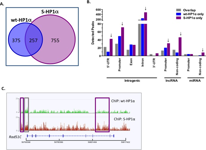
A. Venn diagram of the total binding loci of wild-type and SUMO3-fused HP1α (wt- and S-HP1α) as identified by ChIP-Seq analysis. B. S-HP1α binds more 5′-UTR, promoters, and introns in intragenic regions and regions transcribing ncRNA. Graph represents distribution of detected peaks (binding sites) of wt and S-HP1α at indicated intragenic region. Gray bars indicate binding sites shared by both HP1α types (Overlap) while binding sites unique to wt-HP1α or S-HP1α only are labeled appropriately. Arrows highlight chromosomal regions with a difference between binding profiles of S-HP1α and wt-HP1α. C. ChIP-Seq track at the Rad51C loci that highlights the number and position of the binding sites for wt- and S-HP1α. Purple box indicates a unique S-HP1α binding site while gray box is common to both wt and S-HP1α.