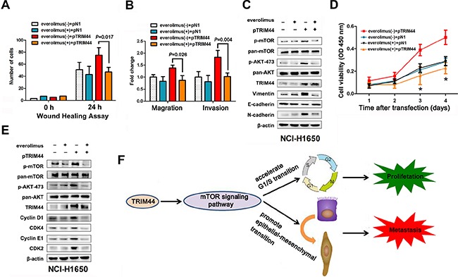
Figure 6. TRIM44 promotes metastasis and proliferation via the mTOR signaling pathway.

NCI-H1650 cells were transfected with p-N1 or p-TRIM44 prior to treatment (or not) with 2 mM everolimus as indicated. (A) Wound healing assays were performed to examine the migration of NSCLC cells. P values were calculated using Student's t-test. (B) Transwell assays were performed to investigate changes in the migratory and invasive ability of NSCLC cells. P values were calculated using Student's t-test. (C) Proliferation of NCI-H1650 cells was measured in a CCK-8 assay at different time intervals (from 24 to 96 h). All values represent the mean ± SEM of triplicate measurements. All experiments were repeated three times, with similar results. *P < 0.05 (Student's t-test). (D) Western blot analysis of components of the TRIM44/mTOR signaling pathway and EMT markers in NCI-H1650 cells. (E) Western blot analysis of components of the TRIM44/mTOR signaling pathway and cell cycle markers in NCI-H1650 cells. (F) Proposed model for TRIM44-induced malignant tumor progression. Increased expression of TRIM44 activates the mTOR signaling pathway, thereby accelerating the G1/S transition and promoting EMT with malignant changes leading to metastasis and proliferation.
