
Figure 6. Induction of GFP signal upon 5 uM cisplatin treatment in GFP- cells.
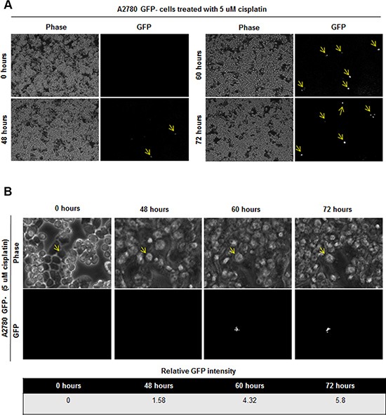
(A) Time lapse imaging of GFP- cells treated with 5 uM cisplatin showed induction of GFP signal in initially GFP–cells. 0.6%, 0.8%, and 1.1% of cells became GFP+ at 48, 60, and 72 hours, respectively. (B) Tracing of one cell which is induced to become GFP+ is shown. Cell of interest indicated with yellow arrow.