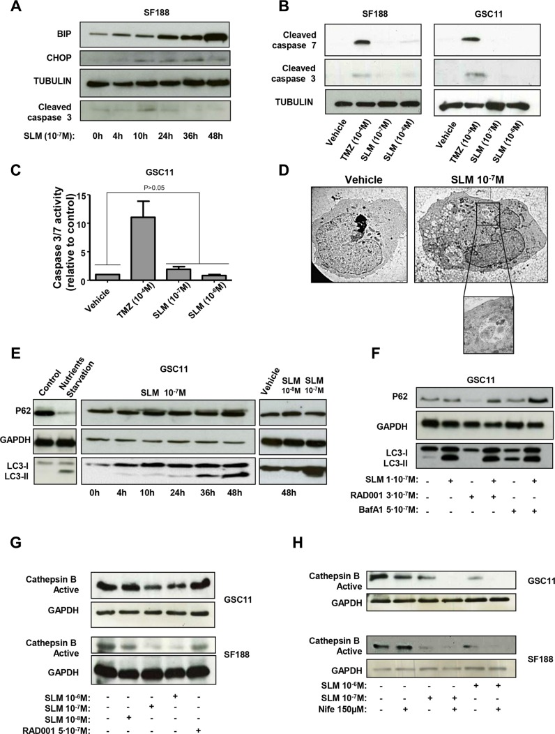
Figure 2. SLM induces ER stress and an aberrant autophagic flux.
(A) SF188 cells were seeded at a density of 1 × 10⁻⁵ cells per well in a 6-well plate. The following day, cells were incubated with SLM (1 × 10⁻⁷ M) and collected at the indicated times. Samples were analyzed by western blot for BiP, CHOP and cleaved caspase 3. α-Tubulin was used as loading control. The western blot shown is representative of three independent experiments. (B) SF188 and GSC11 cells were incubated with SLM (1 × 10⁻⁸ M or 1 × 10⁻⁷ M) or TMZ (1 × 10⁻⁴ M). Cells were collected after 48 h, and samples were analyzed by western blotting for cleaved caspase 7 and cleaved caspase 3. Tubulin was used as the loading control. The western blot shown is representative of three independent experiments. (C) GSC11 cells were seeded at a density of 5 × 10⁻³ cells per well in 96-well plates. The following day, cells were incubated with either TMZ or SLM at the indicated concentration. 48 hours after treatment, caspase −3 and −7 activities were measured with the Caspase-Glo® 3/7 Assay. The results are expressed as mean values ± SD from three independent experiments and caspase −3 and −7 activities in treated cells are represented relative to the corresponding activities in non-treated cells. (D) Transmission electron microscopy analysis. GSC11 cells were treated with SLM (1 × 10⁻⁷ M) and harvested 48 h later. The micrographs shown are representative of the morphologic features observed (1600× magnification). (E) GSC11 cells were incubated with SLM (1 × 10⁻⁸ M or 1 × 10⁻⁷ M) and collected after different incubation times. Samples were analyzed by western blotting for p62 and LC3-I to LC3-II conversion. GAPDH was used as the loading control. The western blot shown is representative of three independent experiments. Nutrient-starved cells were used as a positive control for autophagy. (F) GSC11 cells were seeded at a density of 1 × 10⁵ cells per well in 6-well plates. After 24 h of culture, cells were incubated with RAD001, SLM or bafilomycin A1 (BafA1) at the indicated concentrations. Cells were collected 48 h later and subjected to western blot analyses. The western blot shown is representative of three independent experiments. (G) GSC11 and SF188 cells were incubated with SLM and RAD001 at the indicated doses. Cells were collected 48 hours after treatment, and samples were analyzed by western blotting for active cathepsin B; GAPDH was used as the loading control. (H) GSC11 and SF188 cells were incubated with Nife (1, 5 × 10⁻⁴ M) alone or in combination with SLM (10−7 M or 10−8 M). Cells were collected after 48 h, and samples were analyzed by western blot for active cathepsin (B) GAPDH was used as the loading control. The western blot shown is representative of three independent experiments.

