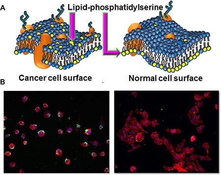
Figure 2. Lipid-PS expression difference between cancer vs normal cells.

(A) Schematic representation of membrane lipid asymmetry in cancer and normal cells and lipid-PS externalization on cancer cells. (B) Staining of HCC4017 (left) and HBEC30KT (right) with PS targeting bavituximab antibody. Only HCC4017 cells express PS.
