
Figure 5. miR-638 inhibits HCC progression in vivo .
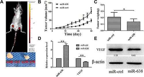
miR-638 and control vector-transfected SMMC-7721 cells were injected subcutaneously into different posterior flanks of the same nude mice. The mice were observed for xenograft growth for 4 weeks. (A) The tumor volume was assessed in situ by a small animal imaging analysis four weeks after tumor inoculation (left flank: ctrl-transfected; right flank: miR-638-transfected). The lower portion of the figure showed the gross morphology of tumors at four weeks after tumor inoculation. (B) Tumor growth curves. (C) Tumor weight. (D) The levels of miR-638 and VEGF mRNA in the tumor tissues from the animals. (E) The protein levels of VEGF in the tumor tissues from the animals. *P < 0.05, **P < 0.01.