
Figure 5. Impact of Notch3 and NAC on HeLa cell malignancy.
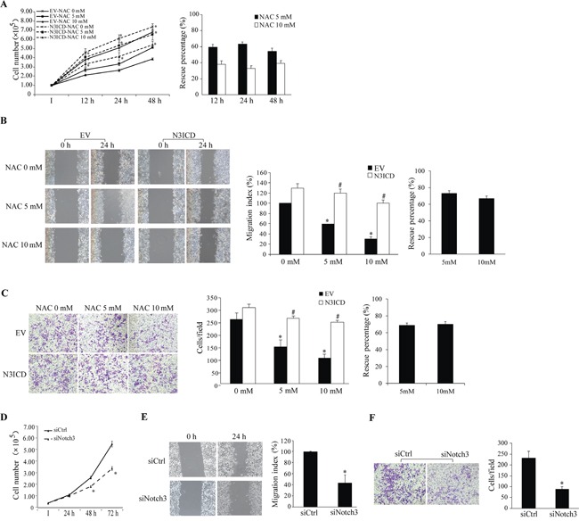
N3ICD overexpression rescues NAC-induced inhibition of proliferation (A), migration (B), and invasion (C). A. Numbers of EV- and N3ICD-transfected cells were counted at 12-48 h after NAC treatment (0-10 mM, left panel). *, p < 0.05 compared with the EV-transfected cells within the same treatment and time point. B. Results of the wound healing assay (left panels) were expressed as the migration index (the distance migrated relative to the initial scraped gap) and that of EV-transfected cells without NAC treatment was set as 100% (middle panel). C. Cells per field on the insert membrane were imaged (left panels) and counted (middle panel). B and C: *, p < 0.05 compared with no NAC treatment; #, p < 0.05 compared with the EV-transfected cells within the same treatment. Percent rescue (A-C, right panels) after N3ICD expression was calculated by dividing the net change after NAC treatment in N3ICD-transfected cells by that in EV-transfected cells. Notch3 siRNA knockdown inhibits cell proliferation D., migration E., and invasion F. as assessed by the same approaches described above. Representative images for migration and invasion were shown. *, p < 0.05 compared with the siCtrl-transfected cells. All data are presented as mean ±SE, n=3. I, the initial seeded cell number. EV, empty vector; N3ICD, Notch3 active intracellular domain; siCtrl, scrambled siRNA; siNotch3, Notch3 siRNA.