
Figure 2. Immunohistochemical and kaplan-meier analyses of intratumoral CD16 and CD8, and the ratio of the two.
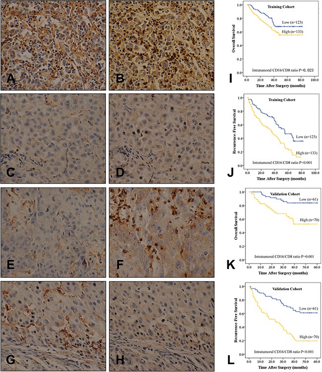
Consecutive sections were used for immunohistochemical staining of intratumoral CD16- (A, C, E, and F) and CD8- (B, D, F, and H) positive cells, which were divided into four subgroups: (A and B) both high; (C and D) both low; (E and F) low CD16 and high CD8 expression; (G and H) high CD16 and low CD8 expression (400× magnification). (I–L) Overall survival (OS, I and K) and recurrence-free survival (RFS, J and L) based on intratumoral CD16/CD8 ratio in HCC patients after curative resection in the training (I and J) and validation cohorts (K and L).