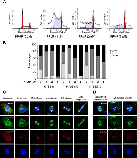
Figure 4. PPMP induces G2/M cell cycle arrest and affects the dynamics of microtubulin.

Flow cytometry analysis of cell cycle was conducted using (A) KYSE510 cells and each of (B) 3 human esophageal cancer cell lines treated for 24 h with the indicated concentrations of PPMP. Cells were harvested and cell cycle was assessed by PI staining. Interphase and mitotic KYSE30 cells treated with (C) DMSO or (D) PPMP were observed by immunofluorescence. β-Tubulin, γ-tubulin, and DNA were stained with anti-β-tubulin (green), anti-γ-tubulin (red), and DAPI (blue), respectively. Merged images are also shown at the top, where co-localization of β-tubulin and γ-tubulin results in a yellow color.
