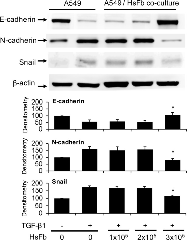
Figure 1. Suppression of A549 EMT by soluble factors of fibroblasts.
A549 cells (1 × 10⁵ cells) were plated at the lower chamber and Hs68 cells (HsFb) at various cell numbers (1~3 × 10⁵ cells) were plated at the upper chamber. Both chambers were co-incubated with or without TGF-β1 (5 ng/mL) for 48 h. A549 cells were harvested and lysed. E-cadherin, N-cadherin and Snail in the cell lysates were analyzed with Western blotting. The upper panel shows representative blots and the lower panel the densitometry. Error bars denote mean ± SEM (n= 3). * indicates P < 0.05 compared to control.

