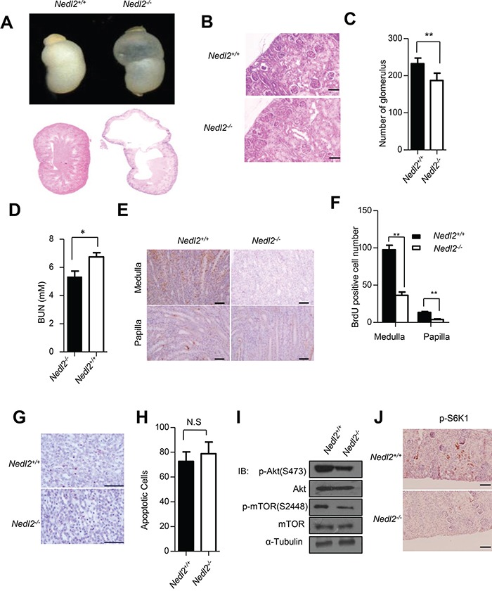
Figure 1. Kidney development defects in Nedl2−/− mice.

A. About 38% (5/13) of Nedl2-deficient newborn mice showed unilateral or bilateral hydronephrosis of distended kidneys (upper panel), HE staining of kidney sections from newborn Nedl2+/+ and Nedl2−/− mutant mice showing severe developmental defect of medulla in Nedl2−/− mice (lower panel). B-C. The presented images (400×) were from Nedl2+/+ and Nedl2−/− mutant mice of the same littermate (B), and the number of glomerular reduced in Nedl2-deficient mice. Statistical analysis of and Glomeruli were counted in the maximal cross-section of each P5 kidney. Five mice were used for each group (C). D. Significant difference of serum level of BUN between Nedl2+/+ and Nedl2−/− mutant mice. (BUN, blood urea nitrogen). E-F. Number of BrdU-positive cells in kidney of Nedl2−/− mice was less than that of in Nedl2+/+ mice, despite medulla or papilla(E). And the values are presented in (F). G. Apoptotic cells were identified by terminal deoxynucleotidyl-transferase-mediated dUTP nick and labeling (TUNEL) staining in kidney of Nedl2+/+ mice and Nedl2−/− mice (P12). No statistical significance existed H. I. Western blot analysis of GDNF/Ret/Akt signaling pathway, fresh kidneys were dissected from P9 mice. J. Significant reduction of p-S6K1 level in Nedl2−/− mice kidney compared with that of Nedl2+/+ mice. Scale bar: 50 μm. Values represent means ± s.d. (*P<0.05, **P<0.01).
