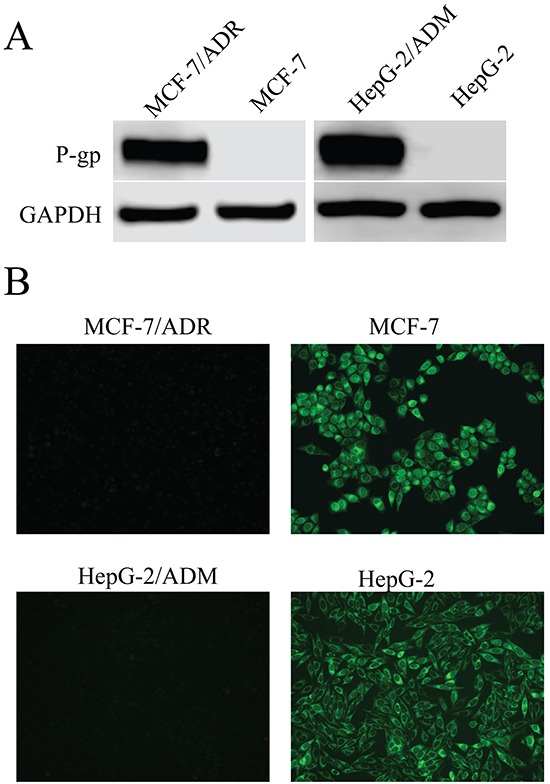
Figure 1. P-glycoprotein (P-gp) expression in MCF-7/ADR and HepG-2/ADM cells in comparison to corresponding parental cell lines, MCF-7 and HepG-2.

A. Western blot analysis of proteins extracted from MDR cells and their parental cells with P-gp antibody. GAPDH was used as loading control. B. Fluorescence microscope detection of the accumulation of rhodamine123 (Rh123) in MDR cells and their parental cells. Images were acquired at 488nm extraction and 535nm emission wavelengths for Rh123.
