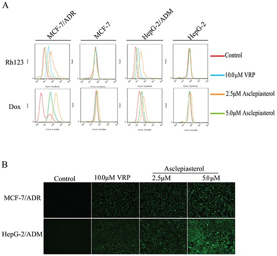
Figure 5. Effect of asclepiasterol at different concentrations on the accumulation of doxorubicin and Rh123.

VRP at 10.0 μM was used as positive control. A. Flow cytometry analysis of the accumulation of Dox (10.0 μM) and Rh123 (10.0 μM) in MDR cells and their parental cells with or without asclepiasterol or VRP treatment. Intracellular fluorescence was analyzed by flow cytometry. B. Fluorescence microscope detection of the accumulation of rhodamine123 (Rh123) in MDR cells after asclepiasterol treatment. Cells were treated with 2.5, 5.0 μM asclepiasterol and 10.0 μM VRP for 48h, and then Rh123 at 10.0 μM was added and incubated for an additional 2h in the dark. Images were taken at 488 nm extraction and 535 nm emission wavelength after washing the cells with cold PBS three times.
