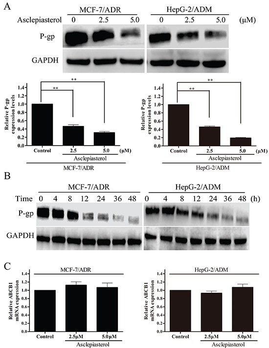
Figure 6. The effect of asclepiasterol on P-gp protein and MDR1 mRNA expression in MDR cells.

A. MDR cells were treated with asclepiasterol at concentrations of 2.5 and 5.0 μM for 48h. P-gp expression was analyzed by Western blotting. GAPDH was used as a loading control. The summary from three independent experiments is shown under the representative blot image. B. Western blot analysis of P-gp expression after a time-course of asclepiasterol treatment. MDR cells were treated with 5.0 μM asclepiasterol for 4 to 48h, P-gp expression was analyzed by Western blotting. GAPDH was used as a loading control. C. RT-PCR analysis of MDR1 mRNA expression in MDR cells after treatment with asclepiasterol at 2.5 and 5.0 μM for 48h. Total RNA was extracted from the cells and the mRNA levels of MDR1 and GAPDH were analyzed by RT-PCR. Independent experiments were performed three times and the summary of the results shown. *P < 0.05, **P < 0.01, compared with the untreated control.
