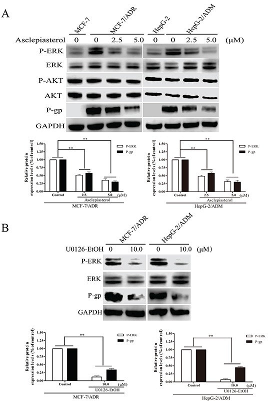
Figure 7. Effect of asclepiasterol on the blockade of ERK1/2 and AKT phosphorylation in MDR cells.

A. Western blot analysis of P-gp expression and ERK1/2, Akt phosphorylation status in MDR cells treated with 2.5 or 5.0 μM asclepiasterol for 48h. GAPDH was used as a loading control. MCF-7 and HepG-2 cells were used as negative controls for detection. B. Western blot analysis of P-gp expression and ERK1/2 phosphorylation status in MDR cells treated with 10.0 μM U0126-EtOH for 12h. Relative P-gp and P-ERK expression of MDR cells from three independent experiments are shown under each Western blot image, *P < 0.05, **P < 0.01, compared with the untreated control.
