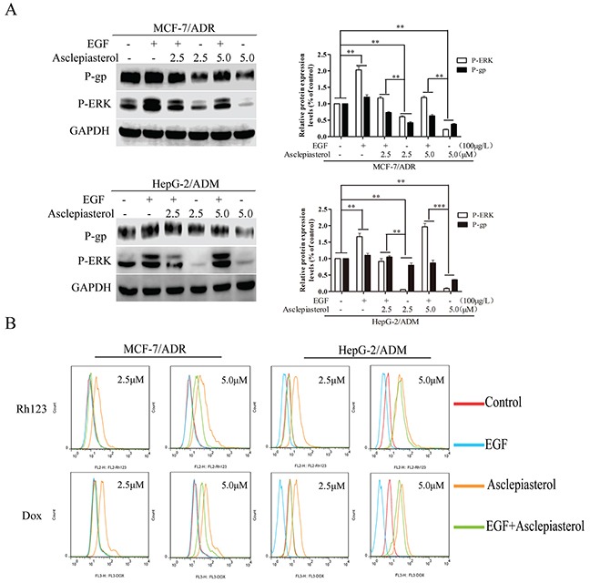
Figure 8. Effect of EGF on asclepiasterol-mediated P-gp down-regulation and P-ERK suppression.

A. Western blot analysis P-gp expression and ERK1/2 phosphorylation after treatment of MDR cells with EGF alone, asclepiasterol alone (2.5, 5.0 μM), or their combination. MDR cells were treated with (+) or without (−) asclepiasterol for 48h to down-regulate P-gp expression on the cell surface and then were treated with (+) or without (−) asclepiasterol combined with 100μg/L EGF for an additional 24h. P-gp and P-ERK1/2 expression were determined by Western blot analysis. GAPDH was used as loading control. Relative P-gp and P-ERK expression of MDR cells from three independent experiments are shown. B. Flow cytometry analysis of the accumulation of Dox (10.0 μM) and Rh123 (10.0 μM) in MDR cells treated with asclepiasterol (2.5, 5.0 μM) in combination with 100μg/L EGF. MDR cells were treated under the same conditions as in (A), and were incubated with Dox (10.0 μM) or Rh123 (10.0 μM) for a further 2h. Intracellular fluorescence was analyzed by flow cytometry. *P < 0.05, **P < 0.01, ***P < 0.001 compared with the untreated control.
