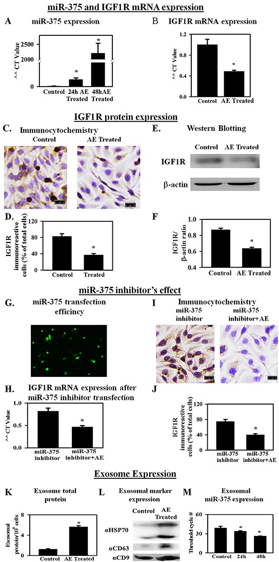
Figure 4. AE increases the expression of miR-375 in ovarian cancer cells as well as in exosomes in the medium.

IGF1R, a receptor for IGF, is associated with proliferation. miR-375 blocks the expression of IGF1R. A. RT-qPCR results showing a >2000-fold increase in miR-375 gene expression (48 h) in SKOV3 cells treated with AE compared to untreated control cells. B. RT-qPCR results (fold change) show that AE reduces IGF1R gene expression in SKOV3 cells at 24 h. C. Immunocytochemistry results show that AE (400 μg/ml) attenuates IGF1R protein expression in SKOV3 cells at 24 h. D. Bar graph shows immunopositive cells as percent of total number of cells counted. E. A representative photograph of SDS-PAGE Western blot analysis of the expression of IGF1R protein in SKOV3 cells treated with AE (400 μg/mL, 24 h). β -actin was used as loading control. F. Bar graph presents IGF1R/β -actin densitometry ratios obtained from 4 independent experiments. G. Representative image of transfected fluorescent anti-hsa-miR-375 miScript miRNA inhibitor in SKOV3 cells at 24 hour. H. RT-qPCR results (fold change) show that AE blocks the effect of transfected miR-375 inhibitor on IGF1R gene expression in SKOV3 cells at 24 h. I. Immunostaining for IGF1R in SKOV3 cells transfected with anti-hsa-miR-375 miScript miRNA with (400 μg/mL) and without AE. J. Bar graph shows IGF1R immuno-positive cells as percent of total cells. K. Total exosomal protein in control and E. officinalis-treated SKOV3 cells (48 h). L. Exosomal marker protein expression in control and E. officinalis treated SKOV3 cells (48 h) culture media. M. MiR-375 expression in exosomes of control and E. officinalis treated SKOV3 cells (48 h). Values are Means ± SEM (n=4), *, P<0.05 compared with control. Bar=25 μm.
