
FIGURE 2.
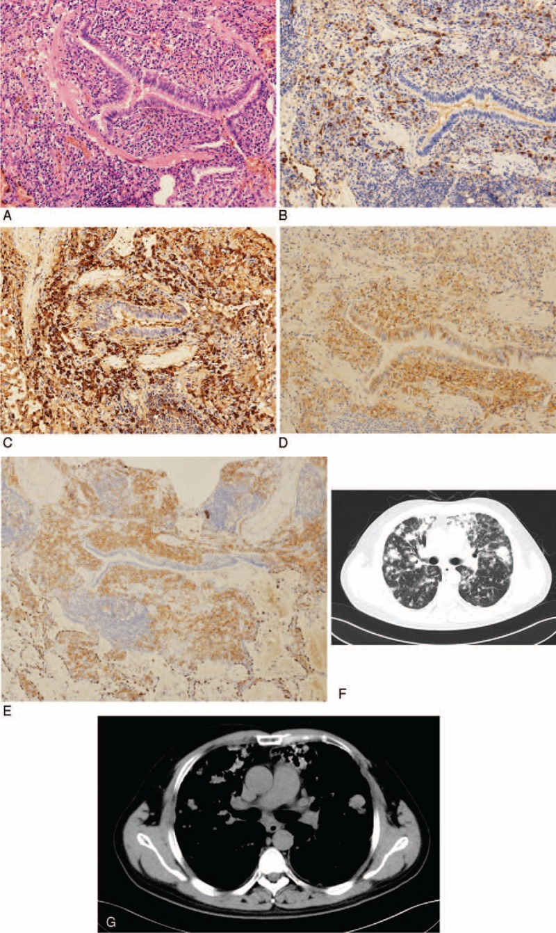
Lung biopsy of a 37-year-old man shows lymphocytes cells infiltration at the peripheral bronchus (hematoxylin and eosin staining, original magnification [A]: ×150). (B–E) Immunohistochemistry shows CD138+, IgG4 (+ >50 per high-power field), IgG4/IgG 46% (B: IgG4 × 150, C: IgG × 150, D: CD138 × 150, E: CD38 × 150). (F and G) Chest computed tomography image of the same man shows diffuse lung parenchymal lesions, including multiple ground-glass opacity and solid nodular manifestations in bilateral lungs.