FIGURE 3.

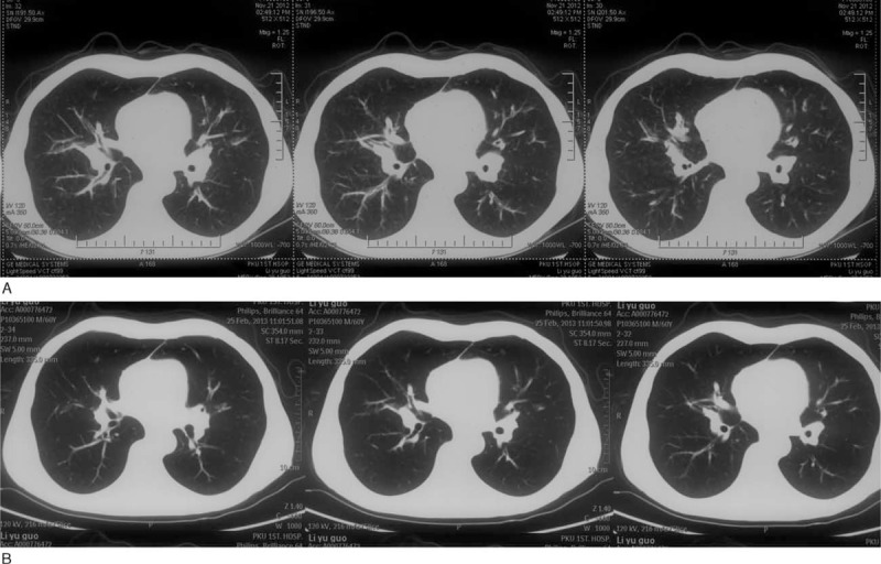
(A) Chest CT image of a 60-year-old man shows bronchial thickening in the bilateral hilar region at diagnosis. (B) Chest CT image of the same man shows obvious improvement after 3 mo treatment. CT = computed tomography.

(A) Chest CT image of a 60-year-old man shows bronchial thickening in the bilateral hilar region at diagnosis. (B) Chest CT image of the same man shows obvious improvement after 3 mo treatment. CT = computed tomography.