
FIGURE 1.
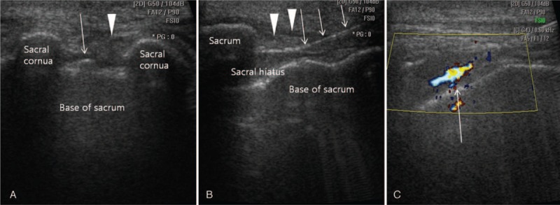
Ultrasound-guided caudal epidural steroid injection. (A) Ultrasound-guided caudal epidural steroid injection in the short axis view. Ultrasound shows the 2 sacral cornus as 2 hyperechoic reversed U-shape structures. The arrowhead is pointing to the sacrococcygeal ligament covering the sacral hiatus. The structure at the bottom is the dorsal bony surface of the sacrum. The arrow indicates the caudal epidural needle. (B) Long axis view showing the needle inside the caudal epidural space. The arrowhead is pointing to the sacrococcygeal ligament. The arrow indicates the caudal epidural needle. (C) Color Doppler Ultrasonography. A predominantly 1-color spectrum is in the caudal epidural space after injection of the contrast media (arrow).