FIGURE 2.

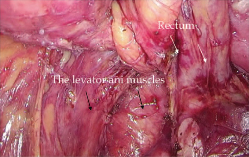
Dissection of the rectum. The rectum (white arrows) was dissected with the TME approach and proceeded circumferentially into the pelvic floor as distal as possible. The levator ani muscles were clearly displayed (black arrows).

Dissection of the rectum. The rectum (white arrows) was dissected with the TME approach and proceeded circumferentially into the pelvic floor as distal as possible. The levator ani muscles were clearly displayed (black arrows).